

- HOME
- Research Content
- Yuji Yokouchi


【Contact】
Developmental Genetics
Institute of Resource
Development and Analysis
Kumamoto University
2-2-1 Honjo, Kumamoto
860-0811, Japan
- TEL :
- +81-96-373-6598
- FAX :
- +81-96-373-6599

Research Content
Project Professor Yuji Yokouchi
Projects
Many medical research groups are attempting to induce cultured stem cells to differentiate into functional cells that are of use in regenerative medicine. Some success has been achieved with respect to hematopoietic, nervous system, and muscular stem cell lineages. To date, however, patients with severe diseases affecting internal organs, such as the liver and pancreas, have not benefited from the use of stem cells largely because the developmental processes of these organs are not fully understood. In this laboratory, we are striving to describe the molecular mechanisms that underlie the normal development of internal organs such as the liver and pancreas, to provide the basic knowledge necessary for regenerative medicine.
1. Histogenesis of the developing chick liver
The liver is the metabolic center of vertebrates and expresses various metabolic enzymes for carbohydrate metabolism and detoxification. In order to perform its functions efficiently, the liver possesses a highly organized and complex tissue structure composed of hepatic parenchymal cells, biliary epithelial cells, and sinusoidal endothelial cells (Fig. 1A). This structure is autonomously organized by intercellular interactions between the embryonic progenitor cells, that is, hepatoblasts, primary sinusoidal endothelial cells, and mesenchymal cells from the septum transversum (Fig. 1B). However, the molecular mechanisms of early liver histogenesis are still unclear. In order to identify and characterize these molecular mechanisms, we have screened the developing chick liver for genes that are expressed during this developmental period. This analysis identified 20 candidate genes. Currently, we are focusing on the epithelial-mesenchymal interactions between hepatoblasts and endothelial cells and are investigating the possible roles of these candidate genes in these interactions.
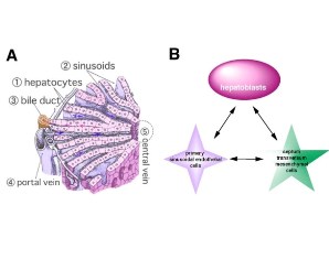 Fig. 1. (A) The microscopic structure of the liver.
(B) The intercellular interactions between the three embryonic precursors in the developing liver.
2. Proximo-distal patterning of the developing chick liver
The liver bud is composed of multi-potent endodermal progenitors. Along the proximo-distal axis, the liver bud gives rise to the ventral pancreatic bud, the extrahepatic bile duct, the gallbladder, and the liver proper (Fig. 2). This pattern of organ development along the proximo-distal axis of the liver bud is conserved in all vertebrates. Investigations of the molecular mechanisms of development of other organs have suggested that patterning involves both region-specific transcription factors and also local organizers that control the region-specific expression of the transcription factors. In order to characterize the molecular mechanisms of patterning in the developing liver, we constructed a fate map of the anterior intestinal portal. We were then able to identify marker genes that are expressed in a region-specific manner in the ventral pancreas, the extrahepatic bile duct, and the liver proper. In parallel with this, we sought to identify the region-specific transcription factors and found that Id3, an HLH-type transcription repressor, is expressed in the liver proper. Id3 is required for the proliferation of hepatoblasts in the developing chick liver. Currently, we are focusing on transcription factors that are expressed in an EHBD-specific manner and are investigating their function and role in the regulation of liver development.
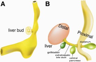 Fig. 2. (A) The liver bud invading into the septum transversum.
(B) The proximo-distal pattering of the developing liver.
3. Asymmetric development of the inner organs
In vertebrates, most organs (for example, eye, limbs, kidney, etc) develop symmetrically, however, some internal organs develop in an asymmetric manner (Fig. 3A). The human congenital disease situs inversus, which causes a reversal of the normal left-right (L-R) asymmetry, suggests that establishment of L-R asymmetry depends on developmental programs coded in genome.
In the developing chick embryo, the establishment of L-R asymmetry is believed to occur as follows: bilateral symmetry first breaks down at Hensen’s node, resulting in the appearance of asymmetric expression of signaling molecules around the node. An inferred Activin-like signal induces a small domain of Bmp-4 on the right side of the node and limits expression of Shh to the left side of the node. Subsequently, broad asymmetric expression of other genes is observed in the lateral plate mesoderm (LPM). The TGF-b family member Nodal is induced throughout the left LPM, where it activates left-sided LPM expression of the transcription factor Pitx2 and thereby affects asymmetric morphogenesis. We previously demonstrated that the Cerberus/DAN family member Caronte (Car), which encodes a BMP antagonist, mediates the transfer of L-R positional information from the node to the LPM. However, we have recently found that Car is not sufficient to explain signal transfer between the node and the LPM because there is a critical gap in this process (Fig. 3B).
Car induces the TGF-b family member Nodal throughout the left LPM, where it activates left-sided LPM expression of the transcription factor Pitx2 and thereby affects asymmetric morphogenesis. However, it is unclear which genes are directly regulated by Pitx2 to achieve asymmetric morphogenesis of internal organs. To identify the genes targeted by Pitx2 and the genes downstream of Nodal, we investigated development-related genes that are expressed in an asymmetric manner prior to asymmetric morphogenesis of the internal organs. We identified four genes that are expressed in an asymmetric manner around the midgut region. Currently, we are investigating the functions of genes that are expressed during development of the pancreas.
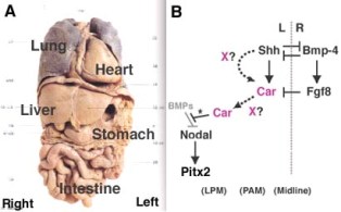 Fig. 3. (A) The L-R asymmetry of the human organs.
(B) The signaling cascades for the establishment of the L-R asymmetry in the developing chick embryo.
